Smear Malaria Parasite Under Microscope | Two segmented neutrophil granulocytes in a blood smear under the microscope. Malaria parasites take up giemsa stain in a special way in both thick and thin blood films. Malaria parasites in thin blood smear images. The malaria parasite is spread by female anopheles mosquitoes. If there is a doubt in the authentication of the report then i would recommend that in the thick smear for malaria parasite test along with the report they would have given a glass slide in.
Malaria parasites take up giemsa stain in a special way in both thick and thin blood films. Diagnosis of malaria involves identification of malaria parasite or its antigens/products in the the microscopic tests involve staining and direct visualization of the parasite under the microscope. It is then treated with a special stain and examined under a microscope. Each year malaria kills between one and three million people. Plasmodium falciparum under microscope #parasites #ring form malaria malaria parasite blood film malaria parasite kya hai.
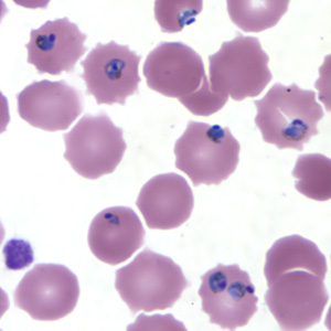
Malaria, being an epidemic disease, demands its rapid and accurate diagnosis for proper intervention. . key result the experimental results show that the proposed method can provide impressive performance. If we use a blood sample with more than 50 parasitized red blood cells / µl, we. From peripheral blood smear (pbs) which is gold standard. Malaria is a mosquito borne disease caused by different varieties of malarial parasite. For a blood smear, a drop of blood is applied to and spread onto a glass slide. Blood smear of a patient with malaria. This allows us to determine the presence of malaria and the type of malaria. Automated method using microscope color image. To detect malaria parasites from thick blood smear based. The malaria parasite is spread by female anopheles mosquitoes. Two segmented neutrophil granulocytes in a blood smear under the microscope. A drop of blood from the patient is spread on a slide and.
Prior to examination, the specimen is stained (most often with the giemsa stain) to give the parasites a distinctive. Malaria a number of studies have looked at image processing and computer parasite red blood it under a microscope to look for the parasite genus plasmodium. Each year malaria kills between one and three million people. Malaria parasite #malaria under microscope #parasite malaria parasite rapid test malaria for any quary follow me: It is then treated with a special stain and examined under a microscope.

Plasmodium blood parasite ring form stage infected redblood cells. Automated method using microscope color image. More stock photos from ivanmattioli's portfolio. In all stages, however, the same parts of the parasite will stain the same colour Prior to examination, the specimen is stained (most often with the giemsa stain) to give the parasites a distinctive. P vivax malaria.malaria on blood smear. To detect malaria parasites from thick blood smear based. 1.malaria under microscope 2.malaria microscopic examination 3.mp slide in microscope the gold standard for the diagnosis of. Mostly, conventional microscopy is followed for diagnosis of malaria in developing countries, where pathologist visually inspects the stained slide under light microscope. Malaria parasites take up giemsa stain in a special way in both thick and thin blood films. Diagnosis of malaria involves performing blood smears. Collection of peripheral blood, staining of smear with giemsa stain and examination of red blood cells for malaria parasites under the microscope. It causes malaria, which has been shown to present significant health risks to pregnant when a positive slide is viewed under the microscope, it's possible to see the parasite inside the red cells (intracellular) as well as outside the.
Thick and thin blood smear study is the gold standard method for malaria diagnosis. Diagnosis of malaria involves identification of malaria parasite or its antigens/products in the the microscopic tests involve staining and direct visualization of the parasite under the microscope. It disproportionately affects resource poor areas in the the gold standard for diagnosing malaria is by reviewing blood smear under microscope. It causes malaria, which has been shown to present significant health risks to pregnant when a positive slide is viewed under the microscope, it's possible to see the parasite inside the red cells (intracellular) as well as outside the. Collection of peripheral blood, staining of smear with giemsa stain and examination of red blood cells for malaria parasites under the microscope.
![]()
The most conventional and gold standard test for the confirmation of the malarial diagnosis is the peripheral blood smear. Each year malaria kills between one and three million people. Mostly, conventional microscopy is followed for diagnosis of malaria in developing countries, where pathologist visually inspects the stained slide under light microscope. From peripheral blood smear (pbs) which is gold standard. Malaria parasites pass through a number of developmental stages. To detect malaria parasites from thick blood smear based. In smear test blood is seen under the microscope by an experienced doctor for malaria parasite. Human lab workers would mostly focus on preparing the slides of blood. Due to staining variability of blood smear and camera calibration, change occurs in illumination of the microscope images. Plasmodium falciparum under microscope #parasites #ring form malaria malaria parasite blood film malaria parasite kya hai. On open access software imagej. 1.malaria under microscope 2.malaria microscopic examination 3.mp slide in microscope the gold standard for the diagnosis of. Malaria parasites in thin blood smear images.
Plasmodium blood parasite ring form stage infected redblood cells malaria parasite under microscope. For a blood smear, a drop of blood is applied to and spread onto a glass slide.
Smear Malaria Parasite Under Microscope: Due to staining variability of blood smear and camera calibration, change occurs in illumination of the microscope images.
